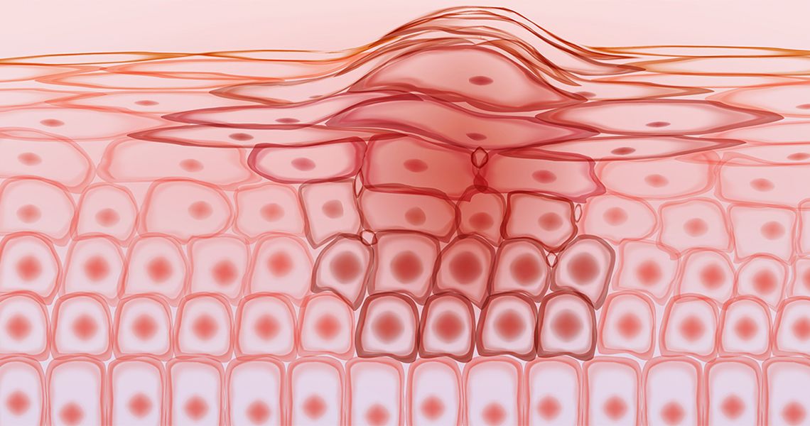
Developed in the 1940s by Dr. Frederic Mohs, Mohs Micrographic Surgery (MMS) is an outpatient procedure that involves surgically removing the visible tumor, along with a thin layer of normal-appearing tissue around and beneath the tumor.
- The moved tissue is then frozen and examined under a microscope. If cancer is seen at the edges or under the surface of the removed tissue, additional tissue is removed from the patient, but only in the area where cancer remains.
- MMS allows for the entire margin of the tumor to be analyzed (called complete peripheral margin analysis) and thus provides the highest cure rates in most skin cancers. The rapid and precise examination of the removed skin reduces guesswork, which means as little healthy tissue as possible is removed. As a result, MMS tends to minimize the size of the surgical wound necessary to remove the cancer completely.
- MMS has become a standard treatment for tumors on the face, hands, and lower legs, where it is important to preserve normal skin, or where healing is difficult.
- MMS is also becoming the preffered treatment of more aggressive tumors such as melanoma and MCC wehre recurrence rates appear to be lower in patients treated with MMS.