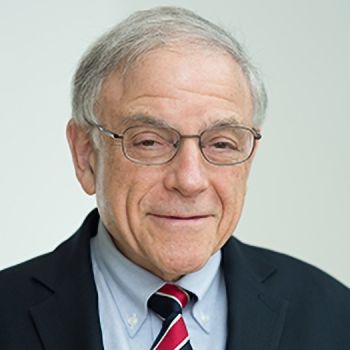
Professor Murray Loew's Laboratory for Medical Imaging and Image Analysis develops new methods for acquiring—and extracting useful information from—medical images. The disciplines involved include pattern recognition, biomedical image and signal processing, and computer vision, with occasional bits of psychophysics and statistics. Although most of the projects deal with images arising in a medical context, his lab’s tools are sometimes applied in other areas.
Areas of active current research include:
Imaging, registration, mosaicking, and analysis of images of paintings: images acquired in visible, x-ray, and infrared (1-2.5 micron) spectral ranges are combined to provide conservators information about underpainting, restorations, and other characteristics. Emphasis is on multi- and hyper-spectral analysis. Super-resolution registration methods are being developed and evaluated. (Supported by NSF)
Hyperspectral imaging for verification of success of RF tissue ablation for treatment of atrial fibrillation (AF). The goal is to provide real-time visualization of tissue damage while performing percutaneous ablation, which will minimize unnecessary tissue injury, and reduce the occurrence of post-ablation AF. (Support from NIH pending)
Development of measures of spatial and temporal salience to guide detection and recognition algorithms, and for detection of anomalies in space and/or time.
Infrared imaging for early cancer detection in the breast: Based on our mathematical modeling of breast thermal and structural characteristics, this work seeks to identify localized temperature increases in the breast arising from an embedded tumor. Infrared imaging is used, and a temperature map derived that will enable objective measurement as well as comparison of the two breasts. Preliminary clinical studies are underway with the GW Breast Clinic.

